Amy Lamb’s professional career spans half a century of science and art. As an undergraduate and graduate student at the University of Michigan, Lamb focused on biology and art history. Her doctoral and post-doctoral research in cellular biology at the University of Michigan, the Basel Institute of Immunology and the National Institutes of Health resulted in the collaborative development of groundbreaking procedures to understand the process of mammalian protein synthesis. Lamb transitioned to art to explore and convey visually the beauty and diversity of plant forms. She perfected her skill in studio photography at Montgomery College, the Corcoran School of Art and the Smithsonian Institution. Lamb approaches photography with a scientist’s precision and an artist’s eye for color and form. Her primary interest is to portray the inherent beauty of plant structure. On a quest to deeply understand her subjects, she is an avid gardener; her indoor and outdoor gardens are her laboratory. Lamb’s work is found in private collections and public institutions such as The National Museum of Women in the Arts, The Baltimore Museum of Art, and The Dallas Arboretum.
How has your training and career as a scientist influenced your artwork, and why did you ultimately choose photography as your medium?
From as early as I can remember art and science have been part of my life. My early education at Kingswood School Cranbrook in Bloomfield Hills, Michigan gave me the opportunity to benefit from a rich integration of science and art. The elegant architecture of Eero Saarinen and the classical sculptures of Carl Milles on the Cranbrook campuses immersed the students in a powerfully influential aesthetic.
In my undergraduate and graduate education, I combined science and art almost every year of study. An internship in electron microscopy kindled my love for photographing the beauty of the internal structures of cells. Through electron micrographs, I could view the delicate, jewel-like structures within cells that keep biological life functioning, from respiration and energy production to the synthesis of proteins.
During my years as a research biologist, I worked primarily at the molecular level where all of the processes and reactions that I studied were invisible to the eye. However, I still envisioned in my mind precise and beautiful images of the molecules, amino acids and nucleic acids, interacting and changing to create new biological products, DNA, RNA and proteins.
A class in photography and a photo experience at the U.S. Botanic Garden opened my mind to an entire new vision and profession. Since the language of vision is accessible across cultural, economic, and educational levels, I could convey to many people the miraculous wonder of nature. My first botanical images of orchids revealed each plant’s uniqueness and diversity. I wanted to reveal the many colors, shapes, and sizes that, together with the plants’ fragrances, enable each plant to attract pollinators, thus ensuring continued existence through reproduction. I have continued now for 25 years to grow, observe and photograph many types of plants. I always seek to share the exquisite beauty of each plant’s character. Each detailed structure, whether a tiny hair or a robust seed pod, is a composite of nature’s flexibility and ingenuity in engineering living, complex and beautiful forms.
Through photography I seek to combine the curiosity, precision, and analytic approach of science with the creativity and flexibility of artistic design. Using powerful lenses that can focus closely on a tiny flower, I want to reveal minute details that are often not easily visible to the naked eye. I photograph in a studio with strobe lighting, positioning each flower, leaf, or stem, almost as if the plant were posing for a portrait. Then I adjust the lighting to reveal tiny structures and textures that highlight the plant’s beauty. When I print my photographs large, the flowers jump out before my eyes. I feel that I am looking into their “soul”, and I can see their unique qualities “face to face.” We are “equals,” and I respect their place, their delicacy and their fragility as well as their stalwart power to fade each autumn and return, with vigor, each spring.
How does cultivating the plant prior to photographing it shape your artistic process?
Part of the passion that I bring to my work is the familiarity that I have with the plants in my garden. I select each plant with care. Initially, I am attracted to a flower because, like a friend, I like it, or I think it is interesting. Then I get to know it. I water it, fertilize it, and try to protect it from predators. And it grows, flowers, attracts, pollinators and produces seeds. Like the Little Prince in Antoine de Saint-Exupéry’s novel, I feel responsible for my flower. This is true for all of the plants in my garden. I feel that I know them. Most likely, in this relationship a year (one life cycle) will pass. This is the time when I will photograph. I want to portray each flower or leaf’s uniqueness. What are the structures that are beautiful and so important to survival? I seek to express the point of view that reveals an unexpected beauty or a surprise structure or the essence of the character of the particular plant. After my photograph is complete, I often press the flowers or leaves, and many weeks later see a new view, like an x-ray, of the dried inflorescence and leaves.
Regarding the specific works that will be on display at Dumbarton Oaks (Stanhopea, Angraecum, and Bulbophyllum), what special steps do you follow when capturing orchids in particular?
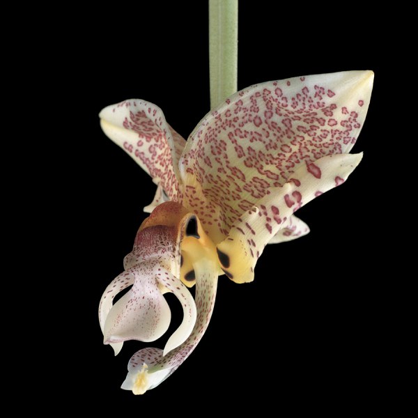
Orchid Study III, S.I. Stanhopea is an amazing plant with many flowers blooming, all cascading downward from the leaves. I wanted to separate the flower and look at its individuality away from the group. I isolated it and portrayed the flower against a black background to highlight its amazing overall structure. Then when I printed it large, I felt vulnerable, just in case this small flower might suddenly grow and gobble me up! In this print the Stanhopea seemed to zoom down with all of its alien structures, like a giant insect leading with its claws. This orchid is so unusual, with its camouflaged petals that remind me of a leopard who will stealthily prowl a forest terrain (fig. 18).
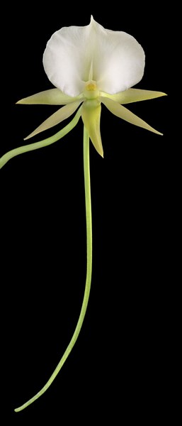
Angraecum II is a star-shaped flower with a very long tail, called a spur. I pondered and tried a couple of ways to photograph this flower, and in the end decided to get very close and use 2 or 3 photographs to obtain detail in the petals and center of the flower and still include all of the lower part of the flower’s “tail” (fig. 5).

Fig. 18. Amy Lamb, Orchid Study III, S.I. Stanhopea (2004).

Fig. 5. Amy Lamb, Angraecum II (2016).

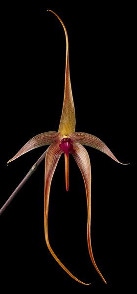
Fig. 19. Amy Lamb, Orchid Study I, S.I. Bulbophyllum (2004).
Orchid Study I, S.I. Bulbophyllum echinolabium is a delicate flower with beautiful transitions of orange and deep magenta. I wanted to capture both the flower’s overall elegance of form and its intricate details. I carefully positioned and lit the flower against a black backdrop to accentuate the graceful shape, crisp edges, and distinct vibrant lines that course through it (fig. 19).